Diastasis Recti Abdominis and Returning to Exercise

Dr. Molly Pierson, PT, DPT • July 16, 2025

Throughout pregnancy, the female body encounters a number of changes to allow room for the baby to grow. Some changes we may notice are that our hips start to tilt forward a little bit more, we have a more extension in our lumbar spine, the rib cage widens to allow room for organs to move up, and, with that, the abdominal wall stretches too. 


With the widening of the abdominal wall, we will also see the connective tissue, the linea alba, in the center of the abdominal muscles widen as well, which is termed
diastasis recti abdominis. 


One fun fact about these changes is that all women during pregnancy have a diastasis recti abdominis, as it allows for adaptations and fetal growth with pregnancy (Gingerich & Prevett, 2023). 


With this particular change, you will hear a lot of talk and concern about pelvic floor weakness, abdominal weakness, and worsening of that widening with abdominal exercises that causes coning or doming of that tissue. However, all of these concerns are not necessarily true.


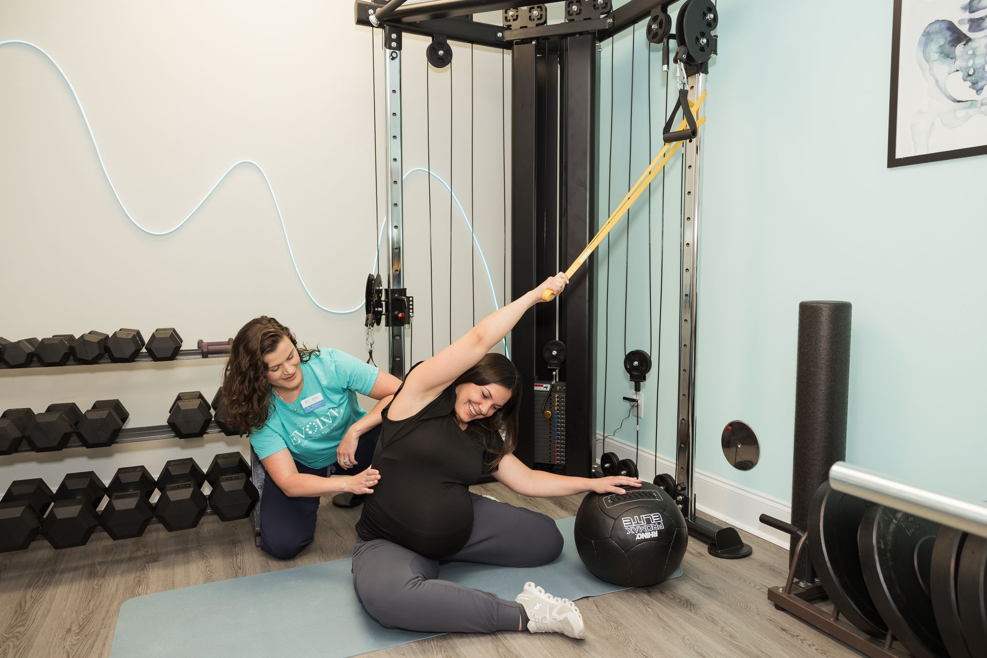
During this blog, we are going to focus on how to return to exercise while addressing your diastasis rectus abdominus or DRA. 


Before we address some of the concerns with DRA, I think it’s important to recognize who this might occur in and factors contributing to it. While the pregnancy and postpartum population is the most common to experience this, it can also be seen in the very lean and muscular athletes, babies, and individuals with increased abdominal circumference (Gingerich & Prevett, 2023). 


There are many factors that can impact the extent of DRA, including genetics, type of delivery, time between and number of pregnancies prior and current activity level, as well as rehabilitation and strength return between pregnancies or before first pregnancy. 


Outside of pregnancy and postpartum and genetics, ability to manage intra-abdominal pressure and willingness to focus on form and quality of movement will also have an impact on an individual's DRA.


So how does this impact our ability to exercising or return to our daily activities? With DRA, many people are aware that coning in the center of the abdomen can cause weakness of the abdominals muscles, particularly rectus abdominis, and pelvic floor weakness as well. 


This is thought to be because coning can worsen our DRA, result in poor management or activation of the core, and difficulty managing intra-abdominal pressure. These theories have led a lot of individuals astray from accessory core work that results in abdominal crunching or rotation of the abdomen to prevent weakness or abdominal/pelvic floor dysfunction. 


However, in a recent study by Gluppe et al., it has been found that there was no increase in prevalence of pelvic floor dysfunction or low back pain among postpartum individuals with DRA compared to those without (2021). It was noted, however, that abdominal weakness and pain were associated with diastasis recti abdominis (DRA) (Gluppe, et al. 2021). 


Along with this, Hills et al., found that individuals who worked on improving abdominal strength with sit ups and abdominal rotation consistently demonstrated improved strength  through abdominal musculature and reduced pain (2018). 


While the inter-rectal abdominal distance may not have improved, working on rotation torque and lower sit ups actually was beneficial with improving the strength of our abdominal muscles regardless of coning (Hills et al., 2018). 


So, collectively the answer to the question above is with progressive loading and creating tension across the abdomen!


Lastly, how do we go about this? If you are returning to exercise and are experiencing DRA, we need to focus on managing our intra-abdominal pressure by creating tension across the linea alba…this means we need to load it with exercise and really focus on muscle activation (Gingerich & Prevett, 2023). 


Ultimately, this means being intentional about our core activation whether we are squatting, picking up our baby, or lifting in the gym. 


And, before you go directly back to kipping and increasing your intensity, take the time to really work on the quality of form and consistency of practice so we can improve our efficiency with movements and reduce risk of injury.


Learn More About Pelvic Floor PT Book a Diastasis Recti Evaluation

References


Gingerich, J. & Prevett, C. (2023, April). CMFA: Pregnancy & postpartum [Powerpoint slide]. Institute of Clinical Excellence.


Gluppe, S., Ellström Engh, M., & Kari, B. (2021). Women with diastasis recti abdominis might have weaker abdominal muscles and more abdominal pain, but no higher prevalence of pelvic floor disorders, low back and pelvic girdle pain than women without diastasis recti abdominis.
Physiotherapy, 111, 57–65. https://doi.org/10.1016/j.physio.2021.01.008


Hills, N. F., Graham, R. B., & McLean, L. (2018). Comparison of trunk muscle function between women with and without diastasis recti abdominis at 1 year postpartum.
Physical therapy and Rehabilitation Journal, 98(10), 891–901. https://doi.org/10.1093/ptj/pzy083


By Dr. Molly Pierson, PT, DPT December 8, 2025
Holly Springs Physical Therapy For Low Back Pain
By Dr. Rebecca Acevedo, PT, DPT September 26, 2025
Physical Therapy for Plantar Fasciitis in Holly Springs, NC
By Dr. Molly Pierson, PT, DPT September 22, 2025
Holly Springs Physical Therapy For Orthopedic Injuries
By Dr. Rebecca, PT, DPT September 10, 2025
Birth Preparation Holly Springs, NC
By Dr. Molly Pierson, PT, DPT September 3, 2025
Typically, once people get into their 3rd trimester of pregnancy, they focus on winding down, resting, and focusing more on stretching. While stretching is helpful, there is more you can do in your 3rd trimester to better prepare for labor, delivery, and recovery after childbirth. Birth prep physical therapy during the third trimester (weeks 28–40) focuses on helping the body prepare for labor and delivery, relieve discomfort, and support postpartum recovery. Here's what it typically includes: Optimize Pelvic Alignment & Mobility Encourages smoother fetal positioning and reduces chances restrictions from your pelvis and surrounding structures. This can include incorporating different positions to help open up different areas of the pelvis, which allow for a smooth transition as the baby moves through the pelvis. Reduce Pain & Discomfort Targets low back pain, pelvic girdle pain, sciatica, and round ligament pain as the baby continues to grow. Diaphragmatic breathing to help manage labor pain and regulate intra-abdominal pressure Prepare Muscles & Tissues for Labor Pelvic floor coordination and perineal mobility can ease labor and reduce tearing risk. Learning to engage abdominal muscles to assist with pushing. Gentle core exercises for transverse abdominis support. Empowerment Through Education Helps you understand pushing mechanics, breathing techniques, and labor positions. This can include: Pelvic Floor Work Focus is on coordination between diaphragm, pelvic floor muscles, and abdominal Learning how to relax the pelvic floor (just as important as contracting it). Gentle perineal massage techniques (usually after 34 weeks). Postural Adjustments & Alignment Body mechanics for daily life (getting out of bed, lifting other children, etc.). Pelvic tilts, cat-cow, and modified squats to maintain mobility. Laboring positions and strategies for fetal positioning Practice labor positions using birth balls, stools, or partner-assisted techniques. Learn strategies for optimal fetal positioning (like Spinning Babies techniques). Manual Therapy Myofascial release, sacral mobilization, and soft tissue work to relieve tension. Addressing hip or SI joint pain. Ultimately, birth prep is tailored to the needs of the mother and her goals when it comes to her birthing experience. The items listed above are just a snapshot of techniques that can be incorporated into sessions to help one become more educated and aware of what the birth experience may encompass, as well as address aches and pains along the way.
By Dr. Rebecca Acevedo, PT, DPT August 14, 2025
Patient Knee Pain Success Story - Physical Therapy Holly Springs, NC
By Dr. Molly Pierson, PT, DPT August 14, 2025
Evolve Physical Therapy in Holly Springs, NC
By Dr. Molly Pierson, PT, DPT August 6, 2025
Dry Needling in Holly Springs, NC
By Dr. Rebecca Acevedo August 1, 2025
There are three primary categories to classify joint mobility: normal mobility, hypermobile, or hypomobile. Hypermobile joints are classified as those that exceed the normative range of motion values. While being extra flexible sounds advantageous, it also requires increased strength and coordination through a larger than usual range of motion. When considering the stability of a joint, there are passive structures such as the shape of the bones, ligaments, joint capsules, labrum and bursa which help the joint maintain its alignment without muscular activation. Dynamic stability of a joint comes from the nervous system working together with our muscles to create smooth and efficient movements. With hypermobile joints, we can work to protect them by improving their dynamic stability through their large range of motion to help reduce risk of injury. Joint hypermobility is also a common presentation in patients with genetic connective tissue disorders such as Ehlers-Danlos Syndromes. Some patients with EDS may experience frequent subluxations, sprains and dislocations due to the instability of their joints. Because of the compromise to their passive stabilizers, sometimes patients with EDS will present with complaints of muscular tightness that truly are stemming from decreased muscular flexibility, as the muscles have been essentially working double time to create stability. A common complaint I often hear from patients is “feeling tight.” But when we check their active and passive range of motion and muscular flexibility, their mobility is above average. Instead, their symptoms of tightness are often caused by muscular weakness, poor endurance, or suboptimal coordination. One treatment strategy I like to try is dry needling with electrical stimulation for pain modulation and improved activation of the targeted muscle’s motor unit, followed by neuromuscular stabilization and strengthening activities to provide long term benefits. Although joint manipulations and stretching feel good at the moment, these treatments are essentially reinforcing excessive mobility in already hyper flexible joints. As Uncle Ben warned Peter Parker (aka Spider Man), “With great power comes great responsibility.” If you have the superpower of being extra bendy, then take into consideration the increased demand on your neuromuscular system to keep your joints stable, and train accordingly! Dr. Rebecca Acevedo, PT, DPT - Holly Springs, NC Physical Therapy
By Dr. Molly Pierson, PT, DPT July 30, 2025
If you’ve ever felt pain in your jaw, heard a clicking sound when you chew, or experienced headaches that seem to start near your ears, you may be dealing with temporomandibular dysfunction, or TMD. This often-overlooked condition affects the temporomandibular joint (TMJ)—the hinge that connects your jaw to your skull and plays a critical role in everyday activities like eating, speaking, and yawning. What Is Temporomandibular Dysfunction? Temporomandibular dysfunction (TMD) is a broad term that refers to disorders affecting the jaw joint and surrounding muscles. The TMJ is one of the most complex joints in the body, allowing both hinge and sliding movements, which allows the jaw to open and close, slide forward/backward, and side-to-side. When something goes wrong with the muscles, ligaments, or joint surfaces, TMD symptoms can develop. Common Symptoms of TMD: Jaw pain or tenderness with opening or closing the jaw Clicking, popping, or grinding sounds when opening or closing the mouth Difficulty chewing or locking of the jaw Headaches and neck pain Facial pain or fatigue What Causes TMD? TMD can be caused by a variety of factors, including: Teeth grinding or clenching (bruxism) Stress and tension leading to muscle tightness Poor posture, particularly in the neck and upper back Injury to the jaw, neck, or head Arthritis or degenerative joint changes Dental issues, such as bite misalignment How Physical Therapy Helps with TMD Physical therapy is a conservative treatment option that can significantly improve TMJ function and reduce pain. A physical therapist trained in TMJ disorders will assess not only your jaw, but also your neck, posture, and muscle patterns to create a personalized treatment plan. Some of the benefits of physical therapy for TMD include: 1. Pain Relief Manual therapy techniques, including soft tissue massage and joint mobilization, can reduce muscle tension and inflammation in the jaw and surrounding areas. 2. Improved Jaw Mobility Therapists use specific exercises to restore normal movement to the jaw, helping to reduce clicking, locking, or restricted motion. 3. Postural Correction Forward head posture and strength deficits of muscles surrounding the spine can put extra stress on the TMJ. PT can help you improve you strength key postural muscles and reduce the strain on your jaw. 4. Muscle Strength and Balance Therapeutic exercises can improve the strength and coordination of the muscles that control jaw movement, reducing dysfunction and recurrence. 5. Stress Reduction Techniques Since stress is a major contributor to jaw clenching and muscle tension, PT often includes relaxation techniques and education on how to manage these habits. Final thoughts If you’re experiencing jaw pain, popping, or limited mobility that lasts more than a couple of weeks, or if you’ve been diagnosed with TMD by your dentist or physician, physical therapy could be a valuable part of your recovery.Temporomandibular dysfunction can have a major impact on your quality of life, but it’s highly treatable—especially when addressed early. Physical therapy offers a safe and effective approach to managing TMD, restoring normal function to your jaw, and helping you return to a pain-free life. References U.S. Department of Health and Human Services. (February 2025.). TMD (temporomandibular disorders) . National Institute of Dental and Craniofacial Research. https://www.nidcr.nih.gov/health-info/tmd